We now analyze the neuron dataset first seen in the comparison of
surface versus direct volume rendering
(Figure 1.4). The dataset is a tomographic
reconstruction of a spiny dendrite on a hippocampal pyramidal neuron
from a rat. The specimen is courtesy of Prof. K. Hama of the National
Institute for Physiological Sciences, Okazaki, Japan. The National
Center for Microscopy and Imaging Research acquired a series of images
of the two micron thick specimen with an intermediate high voltage
electron microscope. The images were processed with single tilt axis
tomography to create a volume of floating point values. This was
quantized to eight bits by histogramming the raw data to determine the
floating point range that best contained the important values.
The histogram volume generated for this dataset has a resolution of
![]() , and was calculated using the Hessian second derivative
measure.
, and was calculated using the Hessian second derivative
measure.
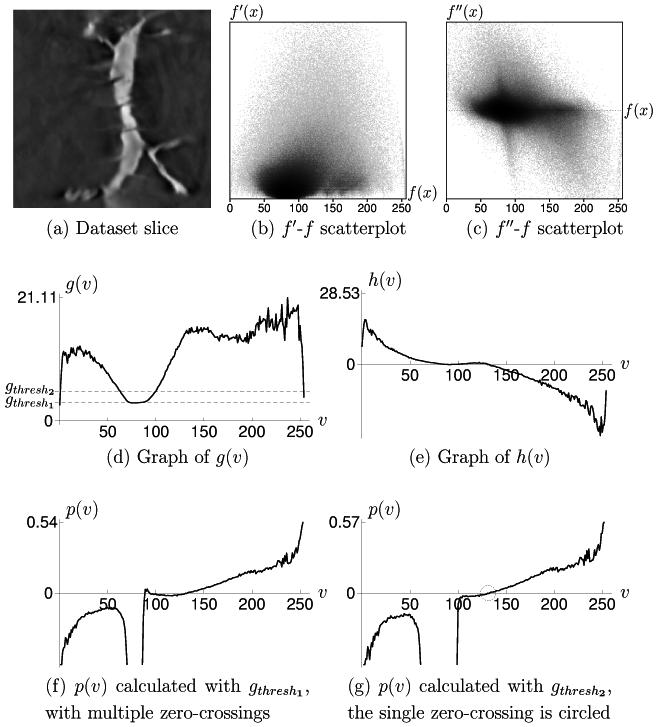
As was mentioned in Section 4.4, the scatterplots
for this dataset (Figures 6.14(b) and
6.14(c)) do not show clear evidence of any boundaries, as
compared to the scatterplots for the other CT datasets analyzed in
previous sections. This is also evident from the graphs of ![]() and
and
![]() (Figures 6.14(d) and 6.14(e)).
Instead of having a noticeable maximum at the data value associated
with the boundary, and being low elsewhere, the predominant feature of
(Figures 6.14(d) and 6.14(e)).
Instead of having a noticeable maximum at the data value associated
with the boundary, and being low elsewhere, the predominant feature of
![]() is its minimum at the data value associated with the
background value (around 80). The graph of
is its minimum at the data value associated with the
background value (around 80). The graph of ![]() is also
atypical-- instead of having one distinct zero-crossing, it lies very
close to the
is also
atypical-- instead of having one distinct zero-crossing, it lies very
close to the ![]() axis along the range of values approximately between
75 to 140.
axis along the range of values approximately between
75 to 140.
We show next the results of two ![]() calculations, based on
different choices for the value of
calculations, based on
different choices for the value of
![]() .
Figure 6.14(d) illustrates how
.
Figure 6.14(d) illustrates how
![]() was chosen to coincide with the average gradient magnitude within the
background value. The resulting
was chosen to coincide with the average gradient magnitude within the
background value. The resulting ![]() (Figure 6.14(f)) has three zero-crossings in the
data value range from 75 to 140, theoretically indicating the
presence of three boundaries in the volume. Yet the cross-section
shows that there are basically just two types of material in the
dataset, background and dendrite, and we are interested in the (single)
boundary between the two. The problem is that the histogram volume
did not succeed in properly measuring the soft boundary that exists
between the dendrite and the background. As a result, straight-forward
application of Equation 5.10 with what appears
to be the correct
(Figure 6.14(f)) has three zero-crossings in the
data value range from 75 to 140, theoretically indicating the
presence of three boundaries in the volume. Yet the cross-section
shows that there are basically just two types of material in the
dataset, background and dendrite, and we are interested in the (single)
boundary between the two. The problem is that the histogram volume
did not succeed in properly measuring the soft boundary that exists
between the dendrite and the background. As a result, straight-forward
application of Equation 5.10 with what appears
to be the correct
![]() did not create a position function
did not create a position function
![]() which is suitable for further analysis.
which is suitable for further analysis.
However, we can choose a
![]() so as to create a position
function
so as to create a position
function ![]() with a single zero-crossing. Raising
with a single zero-crossing. Raising
![]() tends to stretch the graph of
tends to stretch the graph of ![]() away from the
away from the ![]() axis,
eliminating multiple zero-crossings. Using the
axis,
eliminating multiple zero-crossings. Using the
![]() indicated in Figure 6.14(d)
leads to the position function
indicated in Figure 6.14(d)
leads to the position function ![]() seen in
Figure 6.14(g). The new
seen in
Figure 6.14(g). The new ![]() has only one
zero-crossing, which is circled for clarity.
has only one
zero-crossing, which is circled for clarity.
This kind of experimentation with
![]() should be explained,
since it may appear to be somewhat ad hoc. As one can see from the
scatterplot of data value and second derivative
(Figure 6.14(c)), there is a general tendency for the
second derivative to decrease as data value increases. This property
is better represented by the fainter pattern of voxels with very high
and very low second derivatives (located in the upper left and lower
right portions of the scatterplot, respectively), than by the much
larger number of voxels clustered along the region of near-zero second
derivative values. Looking at the second derivative scatterplot, one
can visually interpolate between the faint distribution of voxels with
very high and very low second derivative values in order to find the
approximate zero-crossing of this faint pattern. In our experience,
the single zero-crossing in
should be explained,
since it may appear to be somewhat ad hoc. As one can see from the
scatterplot of data value and second derivative
(Figure 6.14(c)), there is a general tendency for the
second derivative to decrease as data value increases. This property
is better represented by the fainter pattern of voxels with very high
and very low second derivatives (located in the upper left and lower
right portions of the scatterplot, respectively), than by the much
larger number of voxels clustered along the region of near-zero second
derivative values. Looking at the second derivative scatterplot, one
can visually interpolate between the faint distribution of voxels with
very high and very low second derivative values in order to find the
approximate zero-crossing of this faint pattern. In our experience,
the single zero-crossing in ![]() that is revealed by increasing
that is revealed by increasing
![]() is well-correlated with the zero-crossing in the
scatterplot. We now proceed with the second stage of opacity function
generation, by experimenting with different boundary emphasis
functions.
is well-correlated with the zero-crossing in the
scatterplot. We now proceed with the second stage of opacity function
generation, by experimenting with different boundary emphasis
functions.
For an initial
Continuing to make the ![]() peak narrower
(Figure 6.16(a)) leads to an even narrower peak in
peak narrower
(Figure 6.16(a)) leads to an even narrower peak in
![]() (Figure 6.16(b)), and finally the rendering
(Figure 6.16(c)) shows the surface structure of the
neuron. Again, this process was not as automatic as would be ideal,
but to arrive at this rendering (starting with the
(Figure 6.16(b)), and finally the rendering
(Figure 6.16(c)) shows the surface structure of the
neuron. Again, this process was not as automatic as would be ideal,
but to arrive at this rendering (starting with the ![]() calculated
previously) required empirically decreasing only one variable, the
width of the triangular peak in
calculated
previously) required empirically decreasing only one variable, the
width of the triangular peak in ![]() . From here, we can further
decrease the amount of ``haze'' surrounding the neuron by moving the
peak in
. From here, we can further
decrease the amount of ``haze'' surrounding the neuron by moving the
peak in ![]() slightly to the right, to slightly higher position
values (Figure 6.16(d))17. The peak in the new
opacity function moves in the same direction, putting greater
emphasis on higher data values, so as to emphasize the boundary
region closer to the interior of the neuron. The change in the
resulting opacity function is slight (Figure 6.16(e)),
but there is less semi-transparent material surrounding the neuron in
the rendering (Figure 6.16(f)).
slightly to the right, to slightly higher position
values (Figure 6.16(d))17. The peak in the new
opacity function moves in the same direction, putting greater
emphasis on higher data values, so as to emphasize the boundary
region closer to the interior of the neuron. The change in the
resulting opacity function is slight (Figure 6.16(e)),
but there is less semi-transparent material surrounding the neuron in
the rendering (Figure 6.16(f)).